


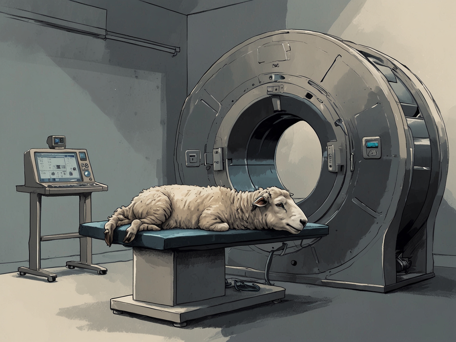
Magnetic Resonance Imaging, or MRI, remains one of the most precise and non-invasive techniques for exploring the inner workings of the brain. The procedure generally requires the subject to remain extremely still to ensure high-quality imaging. For years, sheep, regularly used in neurological research due to the similarities of their brain structure to humans, have undergone MRI scans under general anesthesia. While this approach successfully keeps the animals immobile, it invites a multitude of complications such as stress and potentially dangerous side effects resulting from anesthesia.
© FNEWS.AI – Images created and owned by Fnews.AI, any use beyond the permitted scope requires written consent from Fnews.AI
The latest breakthrough in MRI technology comes from a group of dedicated researchers who have managed to train sheep to complete MRI procedures while awake. This significant advancement aims to reduce stress and most importantly, provide a more accurate representation of brain activity without the distortions induced by anesthesia. The implications of this development are vast and pave the way for more humane and precise methods of studying brain function across various subjects.
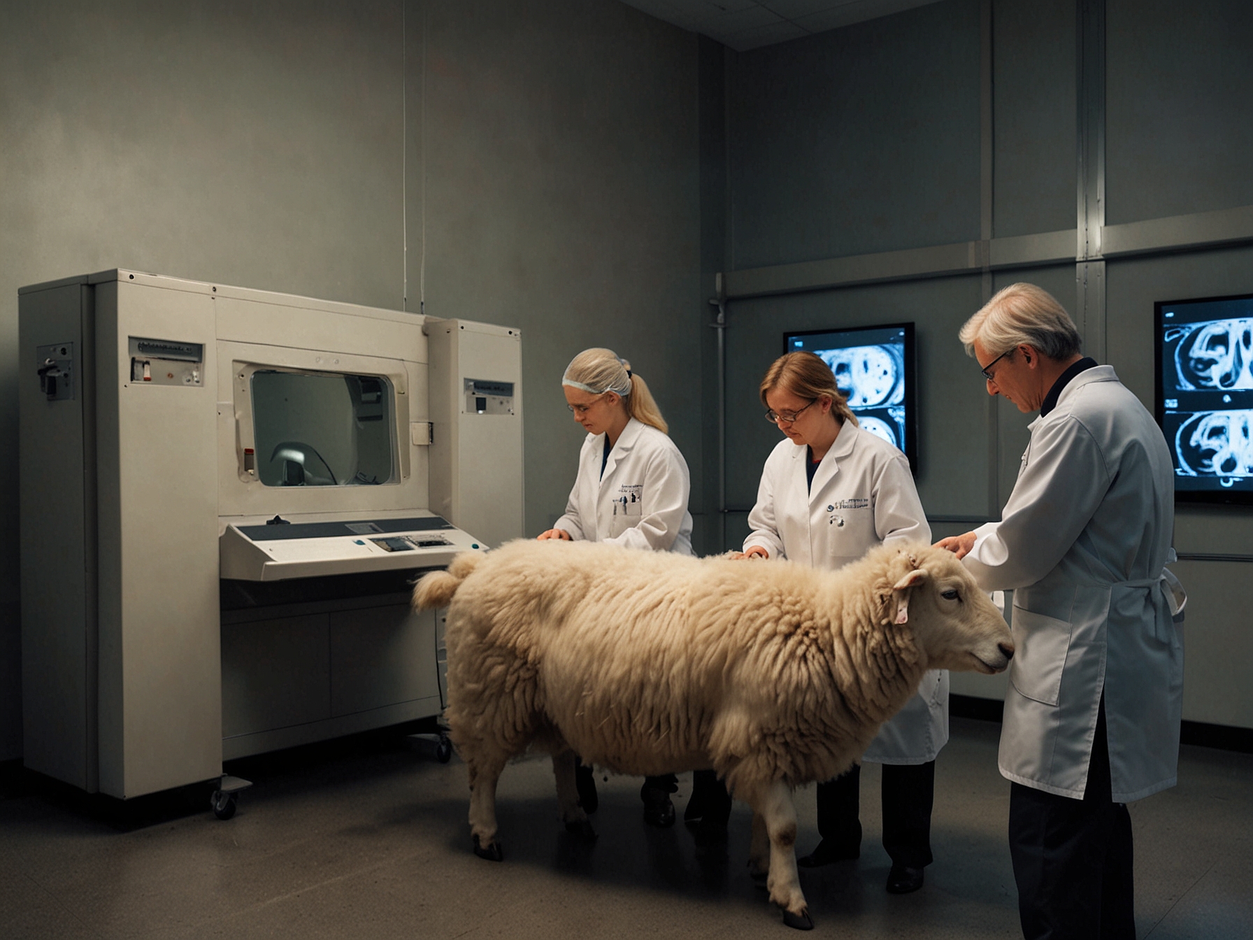
Training sheep for these procedures was no small feat. The first step involved conditioning the animals to enter and remain calm within the confines of the MRI machine. Researchers employed a combination of positive reinforcement techniques, including treats and gentle handling, to encourage voluntary cooperation. Gradually, the sheep were acclimated to the noise and environment of the MRI suite. Over time, they were able to stay almost perfectly still during the scans, providing clear and distortion-free images.
© FNEWS.AI – Images created and owned by Fnews.AI, any use beyond the permitted scope requires written consent from Fnews.AI
This methodology contrasts starkly with the previous use of anesthesia, which, while effective in immobilization, interferes with natural brain activity. Anesthesia can change the functional connectivity of the brain and suppress neural activity, thereby confounding the study results. By eliminating the need for anesthesia, the researchers aim to explore the sheep’s brain activity in a state that is much closer to its natural resting or active state, thereby enhancing the accuracy of the data collected.
One critical component of this training program was to ensure the well-being of the sheep throughout the process. Stress and discomfort were meticulously monitored and minimized, aligning with ethical research practices. The researchers employed advanced behavioral analysis to understand and respond to signs of distress promptly. This sensitivity to the well-being of the subjects is not only ethically imperative but also contributes to the reliability of the data, as stress can significantly alter brain activity.
The successful completion of awake MRI imaging in sheep opens the door to a new era of neurological research. This technique can now be applied to other animals, and potentially even translated to human studies where feasible. It’s worth noting that sheep have been a model for understanding a variety of neurological disorders due to the structural similarities their brains share with human brains. Therefore, the ability to observe brain activity in its natural state offers a significant leap forward in the field of neuroscience.
Furthermore, this advancement may lead to the refinement of MRI procedures beyond academic research. Clinical applications of awake MRI imaging could be seen in veterinary medicine, offering less stressful and more accurate diagnostic tools for a range of animal patients. For example, capturing accurate brain images without the risk associated with anesthesia could markedly improve diagnosis and treatment approaches for neurological conditions in pets.
The reduction in anesthesia use also addresses a critical aspect of the research values underpinning the ‘3Rs’ principle – Replacement, Reduction, and Refinement of animal use in scientific research. By refining the process and removing the need for anesthesia, this technique contributes significantly to the ethical advancement of animal research, illustrating that high-quality data can be achieved without imposing drastic measures on the animal subjects.
Another area of significant interest lies in the potential for longitudinal studies. Awake MRI imaging allows for repeated scanning over extended periods without the cumulative adverse effects of repeated anesthesia. This potential creates avenues for studying the progression of neurological diseases over time within the same subjects, offering unprecedented insights into the disease mechanisms and the efficacy of therapeutic interventions.
Overall, the training of sheep to complete awake MRI imaging marks a celebratory milestone in both neuroscience research and animal welfare. As this practice becomes more widespread, it promises to advance our understanding of brain functions and disorders while adhering to more humane research protocols. The journey from conceptualization to actualization of this technique stands as a testament to the innovative spirit and ethical commitment driving modern scientific research.
Was this content helpful to you?

